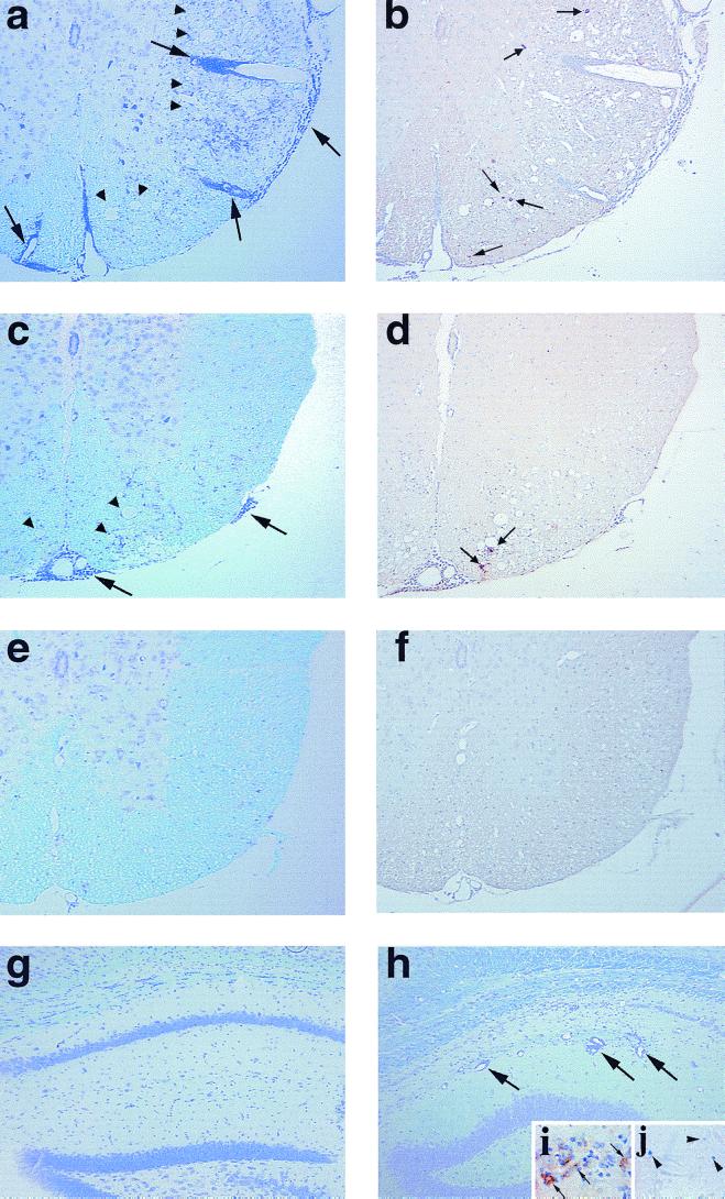
FIG. 6.
Neuropathology during the chronic phase of TMEV infection. Mice were killed 4 months after pDA (a, b, and g), DApB (c and d), or DApBL2M (e, f and h to j) virus infection. pDA virus-infected mice (a) showed severe inflammation (large arrows) with demyelination in the spinal cord white matter (arrowheads) versus DApB virus-infected mice (c). pDA virus-infected mice (b) have more TMEV antigen-positive cells (small arrows) than DApB virus-infected mice (d). In DApBL2M virus-infected mice, spinal cords appeared normal (e) and no viral antigen-positive cells were detected (f). While no lesions were found in the gray matter of the brain in pDA (g) or DApB virus-infected mice, we could see hippocampus inflammation (h, large arrows) in DApBL2M virus-infected mice similar to that of acute disease, even after 4 months postinfection. During the chronic phase of DApBL2M virus infection, both viral antigen (i, diaminobenzidine stain, brown, arrow) and genome (j, BCIP-NBT, blue, arrowhead) could be detected in the pyramidal cell layer of the hippocampus, using immunohistochemistry and in situ hybridization, respectively. (a, c, e, g, and h) Luxol fast blue stain; (b, d, f, and i) immunohistochemistry for TMEV antigen; (j) in situ hybridization for TMEV genome. Magnifications: a to f, ×70; g to j, ×150.

