Fig. 5.
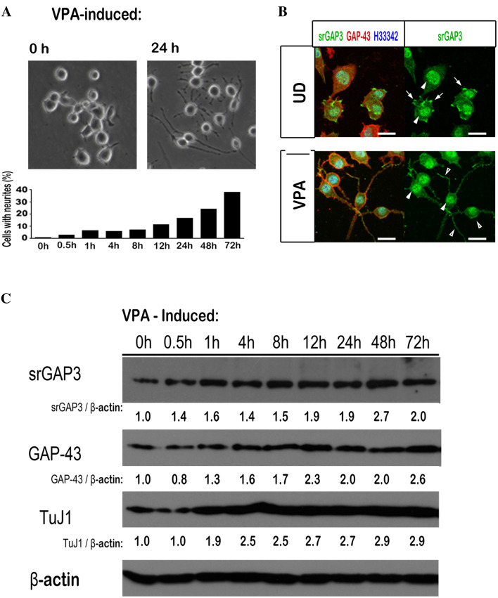
The expression and subcellular localization of endogenous srGAP3 in Neuro2A cells are changed during VPA-induced neuronal differentiation. a Photomicrographs of morphological changes of Neuro2A cells, which are exposed to VPA for 0 or 24 h. The differentiation rates of different VPA stimulation time points are counted and showed in the histogram. b Neuro2A cells cultured in DMEM with 10% FBS (UD, upper panel) or opti-MEM plus 1 mM VPA (VPA, lower panel) are fixed and stained for srGAP3, GAP-43 and DNA. Nuclear-localized srGAP3 is detected both in un-differentiated (UD) and VPA-induced neuronal differentiated Neuro2A cells (indicated by arrowheads). SrGAP3 is also localized at the filopodia and lamellipodia structures before differentiation (indicated by arrows) and at the neurite structures and plasma membrane after Neuro2A differentiation (indicated by open arrowhead). Scale bar = 20 μm. c Lysates from Neuro2A cells exposed to VPA for the indicated times are subjected to western blot analysis. The expression level of srGAP3, together with two other neuronal markers, GAP-43 and TuJ1, is detected. The β-actin is used as a loading control and the relative expression ratio of srGAP3/β-actin, GAP-43/β-actin and TuJ1/β-actin are reported under each lane with 0 h expression level set to 1.0

