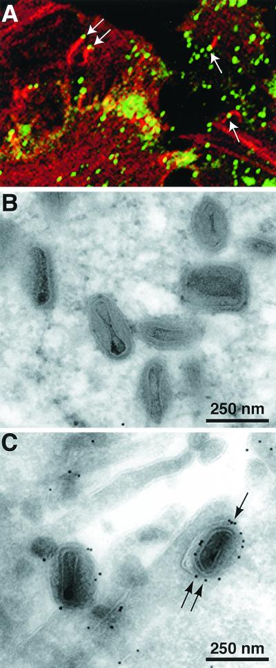
FIG. 2.
Visualization of F13L-GFP by confocal or electron microscopy in cells infected with vF13L-GFP. (A) HeLa cells were infected with 1 PFU of vF13L-GFP per cell. After 18 h, the cells were fixed, permeabilized, stained with rhodamine-phalloidin, and examined by confocal microscopy. Arrows point to green fluorescent particles at the tips of red actin tails. RK13 cells were infected with 10 PFU of vF13L-GFP virus per cell. After 22 h, the cells were fixed, cryosectioned, and probed with rabbit anti-GFP polyclonal antibodies followed by protein A conjugated to 10-nm colloidal gold particles. Electron micrographs show unlabeled IMV (B) and labeled IEV (C). Arrows point to gold grains.

