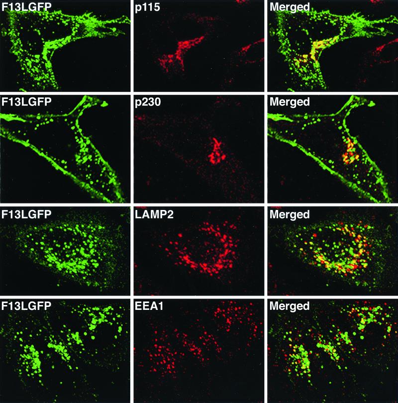
FIG. 4.
Colocalization of F13L-GFP with cellular markers in transfected cells. HeLa cells were transfected with plasmid pF13LGFP for 24 h and then fixed, permeabilized, and stained with the indicated MAbs. Transfected HeLa cells were stained with mouse anti-p115, anti-p230, LAMP2, or EEA1 MAbs followed by rhodamine-conjugated anti-mouse Ig antibody. Stained cells were then examined by confocal microscopy. Green, GFP; red, rhodamine; yellow, overlap of green and red.

