Figure 2.

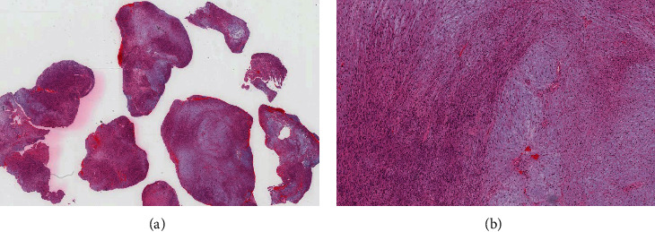
(A) H&E-stained section reveals multiple fragments of lesional tissue with variable cellularity and areas of hemorrhage. (B) Medium-power view showing hypercellular Antoni A “left side” and hypocellular Antoni B “right side” areas.

(A) H&E-stained section reveals multiple fragments of lesional tissue with variable cellularity and areas of hemorrhage. (B) Medium-power view showing hypercellular Antoni A “left side” and hypocellular Antoni B “right side” areas.