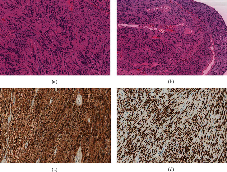
Figure 3.

(A) High-power view of the spindle cells in Antoni A zones of schwannoma that typically show indistinct cytoplasmic borders and elongated, buckled, wavy nuclei embedded in fine, fibrillary, and eosinophilic matrix with characteristic nuclear palisading. (B) A characteristic finding in schwannoma is the presence of single or clusters of irregular vascular channels with hyalinized walls. (C and D) S100 and SOX-10 immunohistochemical stains are diffusely and strongly positive in schwannoma.
