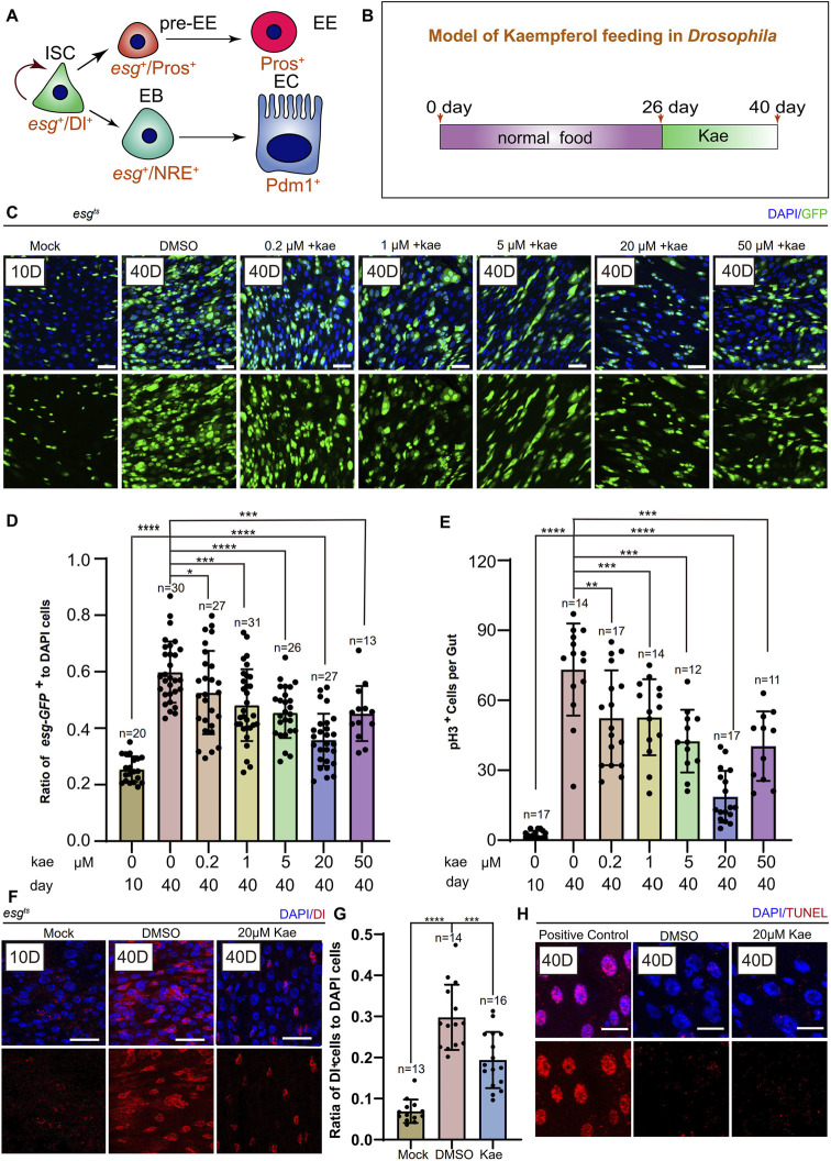
FIGURE 1.
Kaempferol Modulates the hyperproliferation of ISCs in a concentration-dependent manner during aging. (A) ISC division and differentiation model: ISCs (marked as Dl+ and esg+) undergo symmetric division for self-renewal and asymmetric division to produce enteroendocrine progenitor cells pre-EEs (marked as esg+ and Pros+) or EBs (marked as esg+ and NRE+). Pre-EEs differentiate into EEs (marked as Pros+), while EBs develop into ECs (marked as Pdm1+). (B) Model of Kaempferol feeding in aging Drosophila: Mock refers to flies hatched for 10 days and fed a standard diet. Kae indicates flies given Kaempferol. (C) Model of Kaempferol feeding in aging Drosophila: “Mock” denotes flies hatched for 10 days and fed a standard diet. “Kae” refers to flies treated with Kaempferol. (D) The proportion of esg-GFP+ cells to DAPI-stained cells per region of interest (ROI) in Drosophila midguts treated with varying concentrations of Kaempferol (0.2, 1, 5, 20, and 50 µM) compared to untreated controls. (E) The number of pH3+ cells per gut in 40-day-old Drosophila midguts, treated with varying concentrations of Kaempferol (0.2, 1, 5, 20, and 50 µM) or without treatment. (F) Representative images of immunofluorescence staining showing ISCs in Drosophila midguts, treated and untreated with 20 µM Kaempferol. Nuclei stained with DAPI (blue) Dl (red) marked ISCs. The top images represent the merged images and the bottom images represent ISCs. (G) The proportion of Dl+ cells to DAPI+ cells per ROI without or with 20 µM Kaempferol treatment. (H) Representative immunofluorescence images of ISCs in Drosophila midguts, with or without 20 µM Kaempferol treatment. Nuclei are stained with DAPI (blue), and ISCs are labeled with Dl (red). The top images show merged images; the bottom images highlight ISCs. Scale bars denote 25 μm (C, F, H). Error bars show SD. Statistical significance was assessed with Student’s t-tests: *p < 0.05, **p < 0.01, ***p < 0.001; ns indicates p > 0.05.

