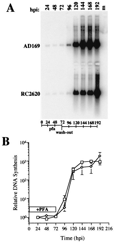
FIG. 4.
Viral DNA accumulation in parental AD169 and mutant RC2620-infected cells in the presence of the DNA replication inhibitor PFA and following the reversal of the PFA block at 72 hpi (wash-out). (A) DNA blot of AD169 and mutant RC2620 viral DNA probed with 32P-radiolabeled pON2260. (B) PhosphorImager analysis of AD169 (□) and RC2620 (◊). Duration of PFA treatment is indicated by an open box (+PFA). Error bars represent standard deviation of the geometric mean of three replicate samples. m, mock-infected DNA.

