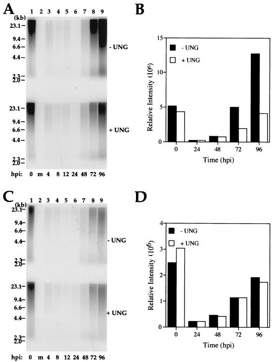
FIG. 6.
Uracil incorporation during parental AD169 and mutant RC2620 virus infection. (A and C) DNA blot analysis of viral DNA isolated from AD169 (A)- and RC2620 (C)-infected HF cells probed with 32P-radiolabeled total viral DNA. Parallel DNA samples from each time point were treated with E. coli UNG (bottom) or left untreated (top). Mock-infected cellular DNA (m) is shown in lane 2 of each blot. Positions of the molecular weight markers are indicated on the left. (B and D) PhosphorImager analyses for selected samples of AD169 (B) and RC2620 (D) viral DNA either treated with E. coli UNG (+ UNG) or left untreated (− UNG) plotted as a function of time.

