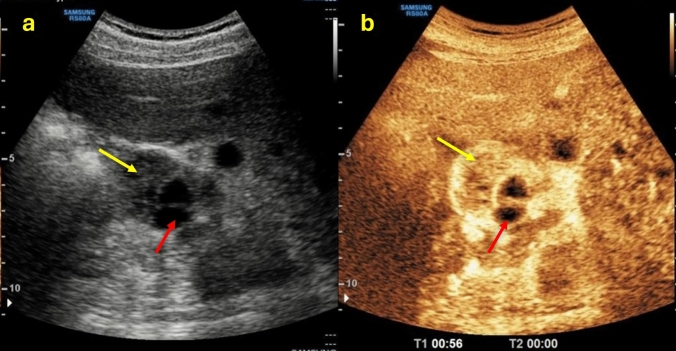
Fig. 1.
(a, b) B-mode US images a showed the presence of a renal mass with a mixed echogenic structure made up of both solid (yellow arrow) and cystic (red arrow) components; CEUS image b showed a better characterization of the mass presenting more than 25% of enhancement of the solid component. The patient underwent a histological biopsy examination that revealed a Grade 1–2 renal cell carcinoma of the clear cell type with diffuse hemorrhagic foci

