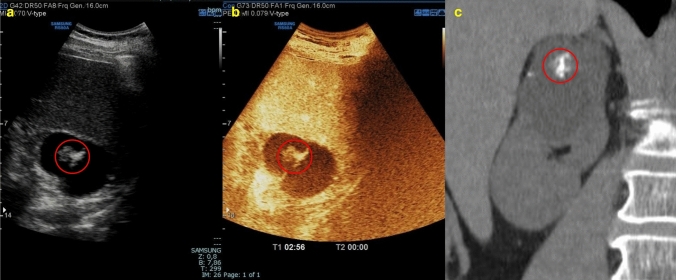
Fig. 6.
(a, b, c) B-mode US image a showed a cystic mass in the right upper pole of the kidney with hyperechoic content due to contextual calcifications (red circle); because of the calcifications, CEUS image b did not allow adequate evaluation of eventual enhancing septa; coronal non-contrast CT scan c confirmed the presence of calcifications but was not able to better characterize the mass. The mass was classified as Bosniak IIF and required further evaluation with MRI

