. 2024 Oct 23;17(10):e261443. doi: 10.1136/bcr-2024-261443

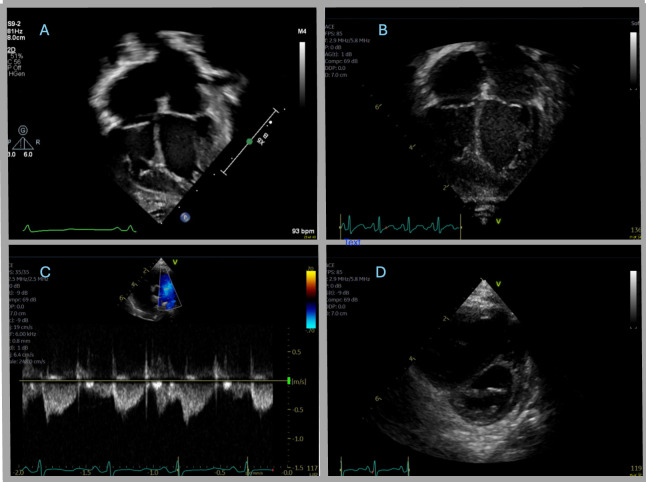
Figure 2. (A) Echo A: apical four-chamber view showing upper normal atrial enlargement with potential prominent trabeculations in the left ventricle myocardium. (B) Echo B: apical four-chamber view showing mild to moderate biatrial enlargement (increased from prior). (C) Echo C: parasternal short-axis view of the ventricles and the interventricular septum that shows flattened end-systolic septal geometry with eccentricity index of 1.4 and septal bounce. (D) Echo C: Doppler pattern tracing of the main pulmonary artery that shows notched pulmonary artery pattern concerning for elevated right-sided heart pressures and potential pulmonary hypertension.

Figure 2