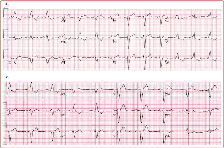
Figure 1: Electrocardiography in Typical and Atypical Left Bundle Branch Block.

A: Twelve-lead ECG showing typical left bundle branch block with QRS duration of >140 ms, and notches in aVL, V4, V5 and V6. B: ECG showing wide QRS and left bundle branch block-like pattern without notches. An electrophysiology study confirmed intraventricular conduction defect with intact Purkinje activation
