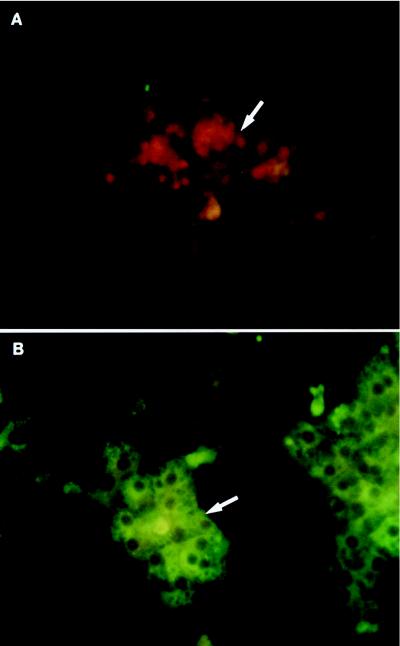
FIG. 3.
Characterization of primary cultures of duck IBDE cells and PDHs by IFA using polyclonal antibodies to duck albumin. Coverslip cultures of primary duck IBDE (A) or PDH cultures (B) were fixed with cold ethanol:acetic acid (3:1) for 5 min at day 2 of culture and processed for IFA using a 1/200 dilution of goat anti-albumin. After reaction with FITC-conjugated anti-goat immunoglobulin G containing Evans Blue, the cells were mounted and viewed. Cytoplasmic fluorescent staining was not detected in the primary IBDE cultures (A, arrow) but was detected in parallel cultures of PDHs (B, arrow). IBDE cells in panel A appeared red because of the Evans Blue counterstain.

