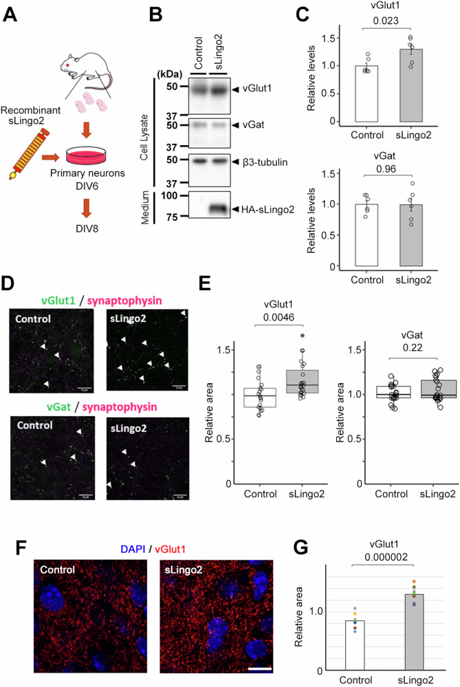
Fig. 4. Synaptic organizer activity of recombinant sLingo2.
A Schematic depiction of this experiment. B Immunoblot analysis of synaptic protein level in primary neuron treated with the recombinant sLingo2. C Quantification of synaptic protein level in B) (n = 6, Mean ± SEM, Student’s t-test, standardized by β3-tubulin). The expression of the vGlut was increased in sLingo2-treated neurons. D Immunocytochemical analysis of vGlut1, vGat, and synaptophysin in primary neurons treated with the sLingo2. Scale bar = 10 µm. E Quantification of vGlut1- and vGat-positive presynapse area in D (n = 21–23, Mean ± SEM, Student’s t-test, standardized by synaptophysin). The relative vGlut1 puncta area was increased by the administration of the sLingo2, but the relative vGat puncta area did not change. F Immunohistochemical analysis of vGlut1 in hippocampal slice cultures treated with sLingo2. Scale bar = 10 µm. G Quantification of mean fluorescent intensity of vGlut1 in F) (n = 8 (control) and 10 (sLingo2) slices from 4, 3 mice each, Mean ± SEM, Student’s t-test).

