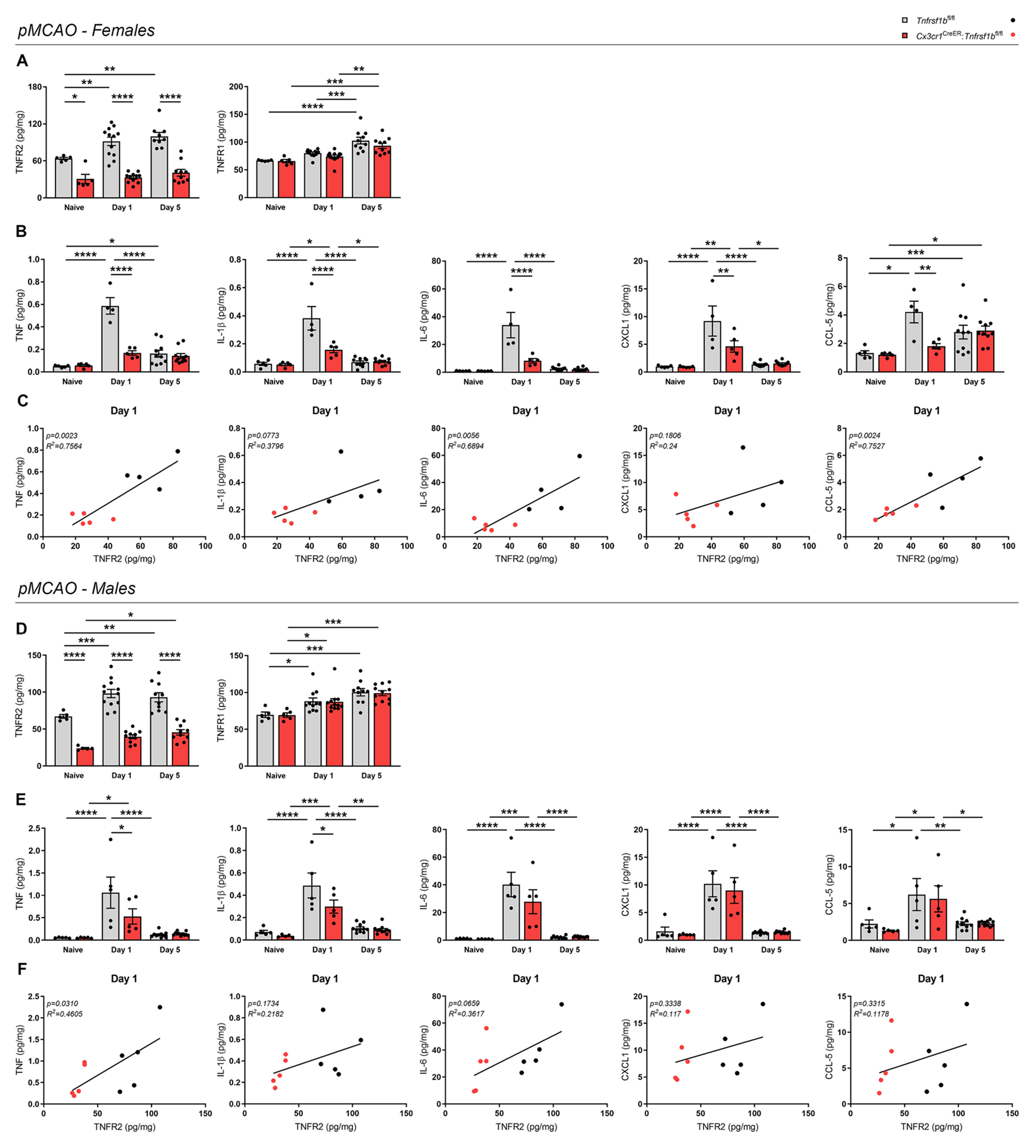
Fig. 3.

Conditional ablation of microglial TNFR2 reduces the protein levels of pro-inflammatory cytokines after ischemic stroke. (A) Quantification of TNFR2 and TNFR1 protein levels in the brain of female Cx3cr1CreER:Tnfrsf1bfl/fl mice and Tnfrsf1bfl/fl littermates in naïve conditions and at day 1 and 5 post-pMCAO (n = 5–12/group). * p < 0.05, ** p < 0.01, *** p < 0.001, **** p < 0.0001; Two-way ANOVA (TNFR2: Interactionns, Time**, Genotype****; TNFR1: Interactionns, Time****, Genotypens) followed by Bonferroni’s multiple comparisons test. (B) Quantification of TNF, IL-1β, IL-6, CXCL1, and CCL-5 protein levels in the brain of female Cx3cr1CreER:Tnfrsf1bfl/fl mice and Tnfrsf1bfl/fl littermates in naïve conditions and at day 1 and 5 post-pMCAO (n = 5–12/group). * p < 0.05, ** p < 0.01, *** p < 0.001, **** p < 0.0001; Two-way ANOVA (TNF: Interaction****, Time****, Genotype****; IL-1β: Interaction****, Time****, Genotype***; IL-6: Interaction****, Time****, Genotype****; CXCL1: Interaction**, Time***, Genotype*; CCL-5: Interaction*, Time***, Genotype*) followed by Bonferroni’s multiple comparisons test. (C) Scatterplot representation of the linear correlation between TNFR2 (x axis) and TNF, IL-1β, IL-6, CXCL1, CCL-5 protein levels (y axis) in the brain of female Cx3cr1CreER:Tnfrsf1bfl/fl mice and Tnfrsf1bfl/fl littermates at day 1 post-pMCAO (n = 4–5/group). For correlation analysis, two-tailed Pearson test was used. (D) Quantification of TNFR2 and TNFR1 protein levels in the brain of male Cx3cr1CreER:Tnfrsf1bfl/fl mice and Tnfrsf1bfl/fl littermates in naïve conditions and at day 1 and 5 post-pMCAO (n = 5–11/group). * p < 0.05, ** p < 0.01, *** p < 0.001, **** p < 0.0001; Two-way ANOVA (TNFR2: Interactionns, Time***, Genotype****; TNFR1: Interactionns, Time****, Genotypens) followed by Bonferroni’s multiple comparisons test. (E) Quantification of TNF, IL-1β, IL-6, CXCL1, and CCL-5 protein levels in the brain of male Cx3cr1CreER:Tnfrsf1bfl/fl mice and Tnfrsf1bfl/fl littermates in naïve conditions and at day 1 and 5 post-pMCAO (n = 5–11/group). * p < 0.05, ** p < 0.01, *** p < 0.001, **** p < 0.0001; Two-way ANOVA (TNF: Interactionns, Time****, Genotypens; IL-1β: Interactionns, Time****, Genotype*; IL-6: Interactionns, Time****, Genotypens; CXCL1: Interactionns, Time****, Genotypens; CCL-5: Interactionns, Time***, Genotypens) followed by Bonferroni’s multiple comparisons test. (F) Scatterplot representation of the linear correlation between TNFR2 (x axis) and TNF, IL-1β, IL-6, CXCL1, CCL-5 protein levels (y axis) in the brain of male Cx3cr1CreER:Tnfrsf1bfl/fl mice and Tnfrsf1bfl/fl littermates at day 1 post-pMCAO (n = 5). For correlation analysis, two-tailed Pearson test was used.
