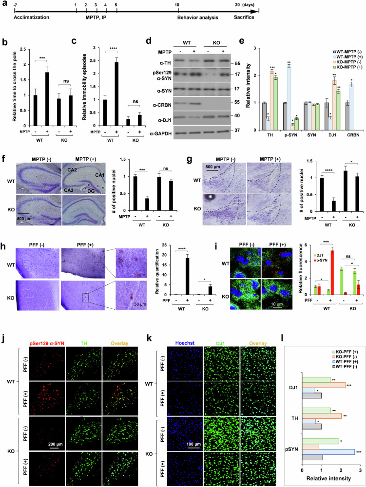
Fig. 6. Crbn-/- mice exhibit resistance towards MPTP- and PFF-induced parkinsonism.
a Schematic representation of MPTP treatment paradigm. b, c Scoring of pole test and tail suspension test for WT and KO mice injected stereotaxically with human WT PFFs (5 μL of 5 μg/μL). d, e Brains were removed from WT and KO mice treated with MPTP, lysates were prepared in RIPA buffer and immunoblotting was performed with the antibodies mentioned. f, g Nissl staining of hippocampus and SNc regions of WT and KO mice injected intraperitoneally with MPTP. CA1, CA2, CA3: cornu ammonis 1, 2, 3 respectively; DG: dentate gyrus. h, i IHC staining of cortex of brains of WT and KO mice injected stereotaxically with human WT PFFs, using DAB substrate for pSer129α-SYN in (h) and alexa-conjugated antibodies in (i). Crystal violet was used for counter staining. IHC staining of SNc (j) and cortex (k) regions of brains of WT and KO mice injected stereotaxically with human WT PFFs with quantification shown in (l). Alexa flour secondary antibodies were used to monitor the levels of TH, DJ1 and p-synuclein. Sections were imaged with an FV1000 confocal laser-scanning microscope. ImajeJ was used for quantification of ROI. Student’s t test and one-way ANOVA was used for the statistical analysis of data.

