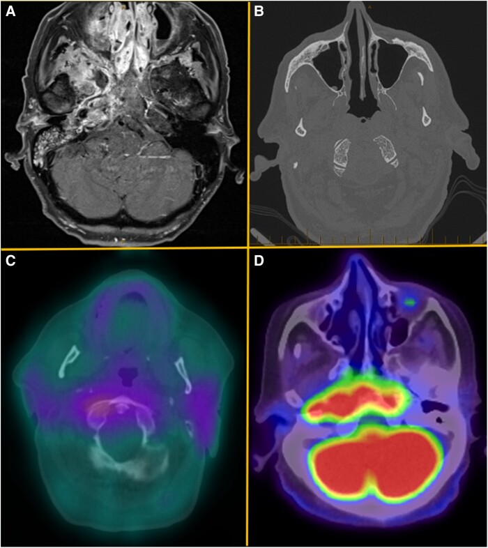
Figure 2.
Medical imaging demonstrating features of central skull base osteomyelitis from selected cases. A (case 1), Axial T1 gadolinium contrast-enhanced magnetic resonance image demonstrating marked enhancement of paranasal sinuses and right skull base involving clivus, sphenoid, and right petrous apex. B (case 9), Axial computed tomographic image demonstrating decortication of the clivus bilaterally along the lateral and anterior margins. C (case 4), Axial fused gallium-67 image showing increased gallium-67 tracer uptake extending posteriorly to the region of the clivus. D (case 3), Axial fused fluorodeoxyglucose (FDG) positron emission tomography image showing intense FDG uptake eroding the clivus. On the right, FDG uptake extends laterally, eroding the petrous part of the temporal bone and involving the foramens lacerum and jugular foramen.

