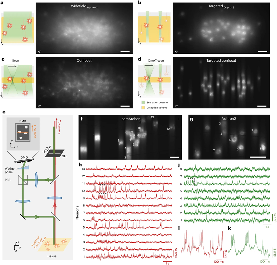
Figure 1. Principle and design of TICO microscope.
(a-d) Illustration (left panel, x-z view) and example images (right panel, x-y view) of different excitation and detection strategies applied to in vivo voltage imaging of Voltron2: (a) standard widefield microscopy where all neurons in a volume are uniformly illuminated and detected with a widefield camera; (b) targeted illumination microscopy where only selected neurons within the focal plane are illuminated, but fluorescence from the entire volume is detected; (c) confocal microscopy where all cells within the volume are illuminated but only fluorescence near the focal plane is detected; (d) TICO microscopy where only selected cells within the focal plane are illuminated, and only the generated fluorescence near the focal plane is detected. Note that images in (a,b) were acquired using a confocal slit size of 156 μm, which produces higher contrast than a true widefield microscope (see Supplementary Fig. 4).
(e) Simplified schematic of TICO microscope (see Extended Data Fig. 1d for a more detailed schematic).. Note that in the actual setup, the slit is fixed and scanning is performed with galvanometers. PBS, polarizing beamsplitter.
(f-k) Demonstration of in vivo voltage imaging of somArchon (f,h,i) and Voltron2 (g,j,k) in the neocortex using TICO microscopy. (f,g) Averaged fluorescence images from somArchon and Voltron2. (h,j) Fluorescence traces from the active neurons labeled in (f) and (g) respectively. (i,k) Zoomed-in fluorescence traces from the boxed regions in (h) and (j) respectively. Scale bars in (a-d, f, g) are 50 μm.

