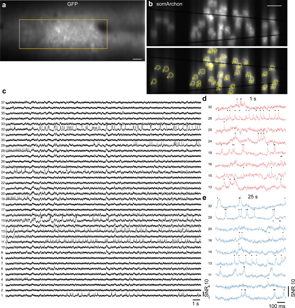
Extended Data Fig. 6. Large-scale imaging of somArchon fluorescence from 37 cells near the visual cortex.
(a) Confocal image of GFP fluorescence. Yellow square indicates actual somArchon imaging FOV shown in (b). Scale bar, 50 μm. (b) SomArchon fluorescence image with 37 cells targeted. Scale bar, 50 μm. (c) SomArchon fluorescence traces of 37 cells over a continuous 30 s recording. Recording speed 775 Hz, imaging depth 100 μm. (d,e) Zoomed-in fluorescence traces of active neurons during 1 s and 25 s of the recording.

