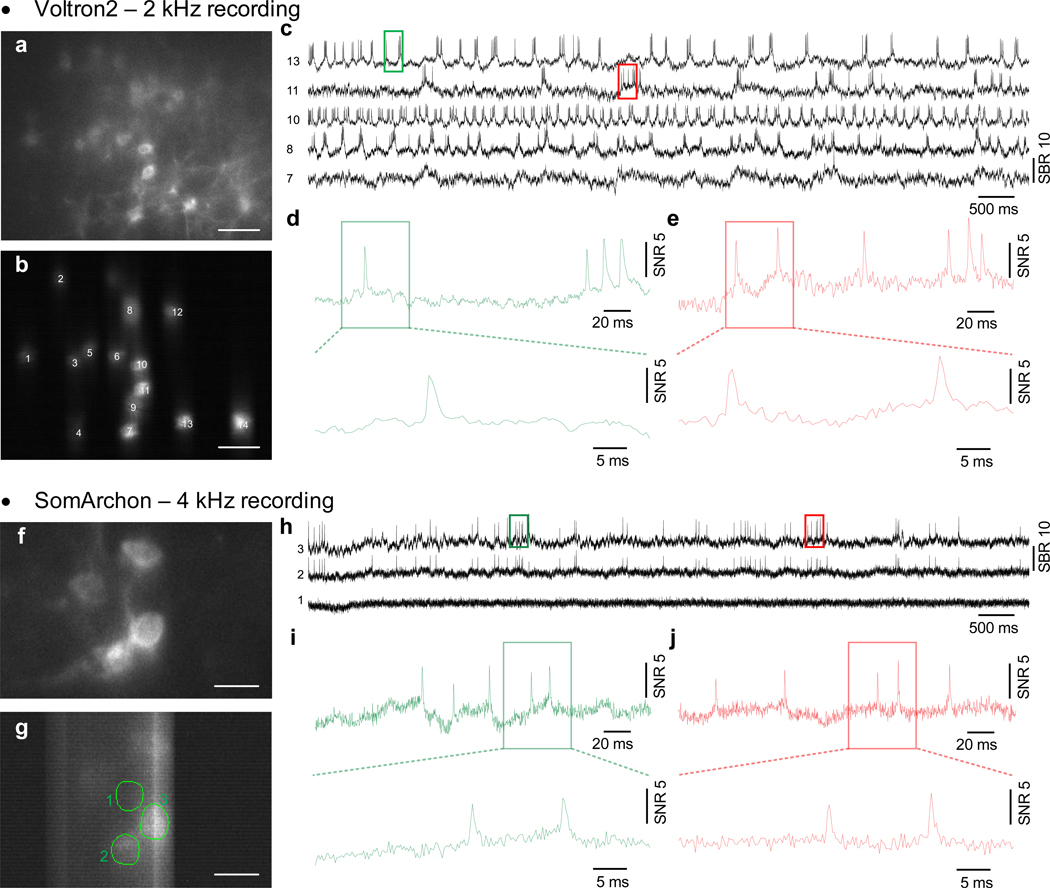
Extended Data Fig. 4. High-speed voltage imaging at 2 kHz and 4 kHz frame rates.
(a) Confocal image of Voltron2 fluorescence. Scale bar, 50 μm. (b) Averaged Voltron2 fluorescence image with 14 neurons targeted within the FOV. Scale bar, 50 μm. (c) Voltron fluorescence traces of 5 active neurons over a 10 s recording. Recording speed 2 kHz. (d,e) Zoomed-in fluorescence traces over the rectangular labeled regions in (c). (f) Confocal image of GFP fluorescence. Scale bar, 20 μm. (g) Averaged somArchon fluorescence image with 4 neurons targeted within the FOV. Scale bar, 20 μm. (h) SomArchon fluorescence traces of 2 active neurons over a 10 s recording. Recording speed 4 kHz. (i,j) Zoomed-in fluorescence traces over the rectangular labeled regions in (h).

