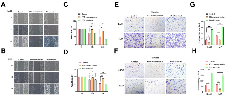
Figure 3.
The impact of FGA on HCC cell migration and invasion. (A, B) Representative images were obtained from the wound healing assay, and the corresponding quantitative analysis of the results of the HepG2 (C) and Huh7 (D) wound healing assays demonstrates the effects of FGA overexpression and knockout in cell migration. (E, F) Representative images were acquired from the Transwell assay, and the corresponding quantitative analysis of the results of migration (G) and invasion (H) in the Transwell assay illustrate the impact of FGA overexpression and knockout on cell migration. (mean ± SD, n = 3; *P < 0.05, **P < 0.01, ***P < 0.001, compared with the control group. Scale: 100 μm).

