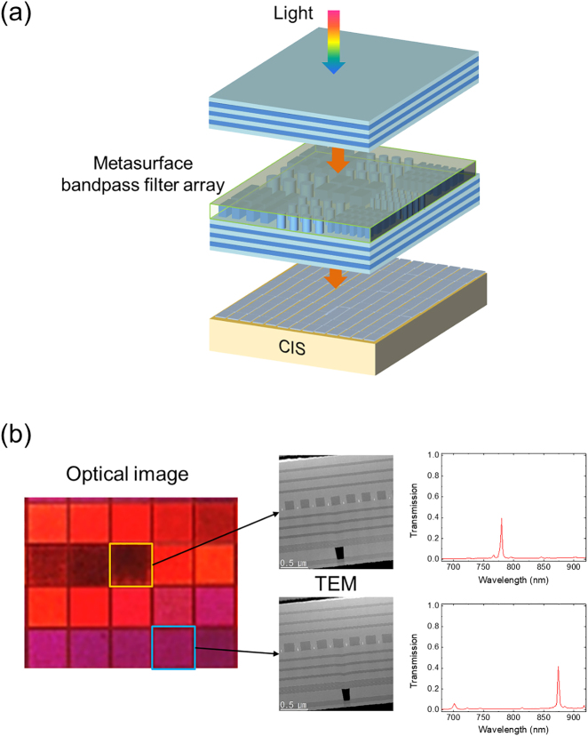
Figure 1:
Meta-spectral imager. (a) Schematic of the meta-spectral imager. (b) Optical microscope image of one pixel made of 20 channels (left). Cross-sectional transmission electron microscope (TEM) images of the two channels (center). The measured transmission spectra of the two selected channels (right).

