
FIGURE 2.
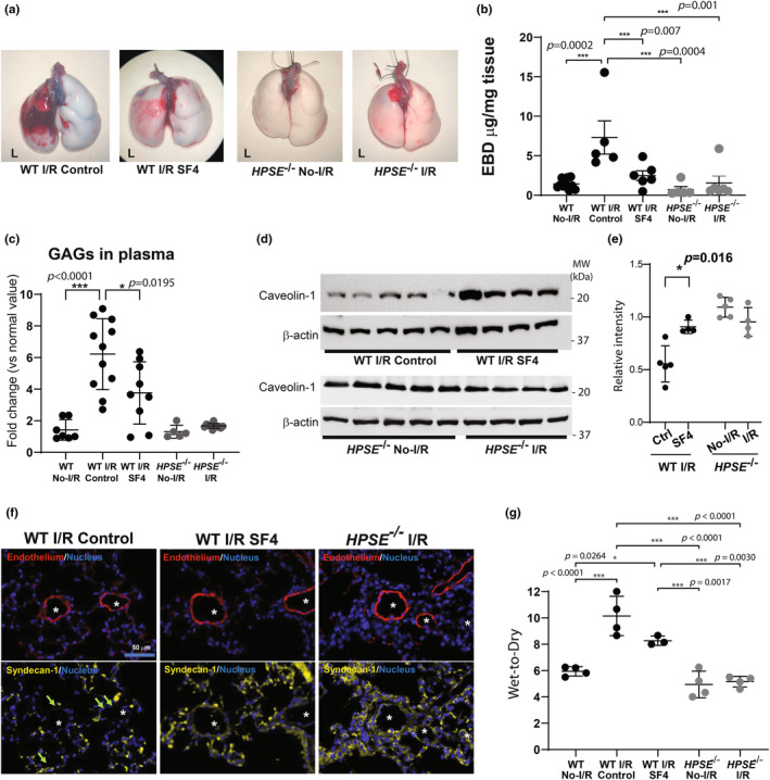
HPSE inhibition or deletion reduces endothelial dysfunction in the lungs after I/R injury. Endothelial barrier function in the lung after ischemia–reperfusion (I/R) injury was evaluated by (a) gross assessment of the lungs after Evans blue dye (EBD) administration and (b) quantitative measurement of EBD extracted from the lung tissue. (c) Plasma glycosaminoglycan (GAG) levels in wild type (WT) and HPSE −/− mice after lung I/R injury were determined using 1,9‐dimethylmethylene blue (DMMB) assay and normalized using the plasma level in uninjured mice of each genotype to calculate fold change. (d, e) Western blotting analysis for caveolin‐1 with β‐Actin as an internal control, in lung tissue after 1 h ischemia/4 h reperfusion and their quantitative analysis (cav‐1/ β‐Actin) (e). (f) Representative images of the immunofluorescent staining for endothelial cells (CD31), syndecan‐1, and nuclei in lung tissue after 1 h ischemia/4 h reperfusion. Asterisks indicate the endothelial lumen. Arrows indicate loss of syndecan‐1 in the endovascular lumen. (g) Wet‐to‐dry ratio of lung tissue after 1 h ischemia/4 h reperfusion. *p < 0.05, ***p < 0.001. No‐I/R, uninjured lungs; I/R, lungs after 1 h ischemia/4 h reperfusion; SF4, mice pretreated with HPSE inhibitor followed by I/R.