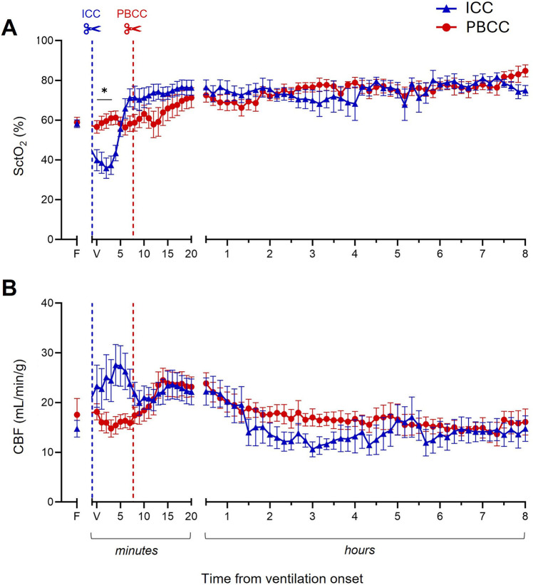
Figure 4.
Cerebral oxygenation and carotid blood flow (CBF) during the 8-h neonatal ventilation. Cerebral tissue oxygen saturation (SctO2, A) and CBF (B) measured over the 8-h ventilation period in lambs with a diaphragmatic hernia (DH) receiving immediate cord clamping (ICC, blue triangles) or physiologically based cord clamping (PBCC, red circles). Timing of umbilical cord clamping relative to ventilation onset shown by blue (ICC; cord clamped prior to ventilation onset) or red (PBCC; cord clamped when a tidal volume of 4 ml/kg was achieved or at a maximum of 10 min) dotted line. Data are expressed as mean ± SEM. In the ICC DH lambs, n = 9 at ventilation onset and n = 4 at 8 h, whereas in the PBCC DH lambs, n = 12 at ventilation onset and n = 4 at 8 h. Mixed-effects analysis with Holm–Sidak multiple comparisons, *p < 0.05 for the effect of group (ICC vs. PBCC). Area under the curve with t-test shown in bar graph where *p < 0.05 for ICC vs. PBCC over the 8 h. F = fetal measurement before ventilation onset, V = ventilation onset.

