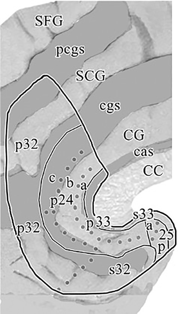
Figure 1.

“Flat map” of the medial surface of the human ACC showing the distribution of subregions and areas from a histological assessment based on a flattening procedure to allow a more accurate visualization of the examined cingulate areas as described in the text. The sulcal depths are noted with a homogeneous gray color. CC, corpus callosum; CG, cingulate gyrus; SCG, superior cingulate gyrus; SFG, superior frontal gyrus; cas, callosal sulcus; cgs, cingulate sulcus; pcgs, paracingulate sulcus. Reprinted with permission from 'Cytology and Receptor Architecture of Human Anterior Cingulate Cortex' by Palomero-Gallagher et al., doi 10.1002/cne.21684, Copyright © 2008 Wiley-Liss, Inc.
