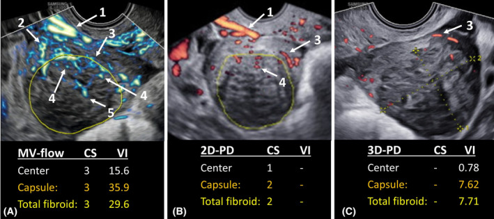
FIGURE 2.

Representative sagittal ultrasound images of one single fibroid obtained with (A) microvascular flow (MV‐flow) imaging; (B) two‐dimensional power Doppler (2D‐PD); and (C) three‐dimensional power Doppler (3D‐PD). Reported outcomes are: Color socore (CS) obtained by MV‐flow or 2D‐PD; and vascular index (VI) obtained by MV‐flow or 3D‐PD. Visible vascular structures are: (1) uterine vascular arcade, consisting of arcuate arteries, (2) radial arteries, (3) fibroid's pseudocapsule, (4) vascular branches penetrating the fibroid, (5) central vascularization.
