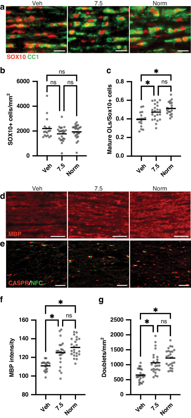
Fig. 3. Clemastine treatment at the minimum effective dose rescues oligodendrocyte differentiation and myelination in the optic nerve at P14.

a Representative images of staining for mature oligodendrocytes (CC1+ cells) and all oligodendrocyte lineage (SOX10+) cells in longitudinal sections of optic nerve at P14 in each treatment condition. Magnification: 20X; scale bars, 20 um. b Column scatter plot of SOX10+ cell density by treatment condition; data are compared by Kruskal–Wallis test. c Column scatter plot of proportion of mature (CC1+) oligodendrocytes out of all oligodendrocyte lineage (SOX10+) cells. *p < 0.05, one-way ANOVA followed by post hoc Tukey’s tests. d Representative images of staining for myelin basic protein (MBP) in P14 optic nerves. Magnification: 20X; scale bars, 30 um. e Representative images of staining for Caspr and neurofascin (NFC) to identify nodes of Ranvier in P14 optic nerves. Magnification: 40X; scale bars, 10 um. f Column scatter plot of normalized MBP intensity in P14 optic nerves. *p < 0.05, one-way ANOVA followed by post hoc Tukey’s tests. g Column scatter plot of density of nodes of Ranvier (identified by NFC/Caspr doublets) in P14 optic nerves. *p < 0.05, one-way ANOVA followed by post hoc Tukey’s tests. For all graphs, horizontal lines depict mean of data sets; ns not significant. For all panels, hypoxia-exposed animals were: Veh (vehicle-treated) and 7.5 (clemastine 7.5 mg/kg/day); Norm, normoxia-exposed animals.
