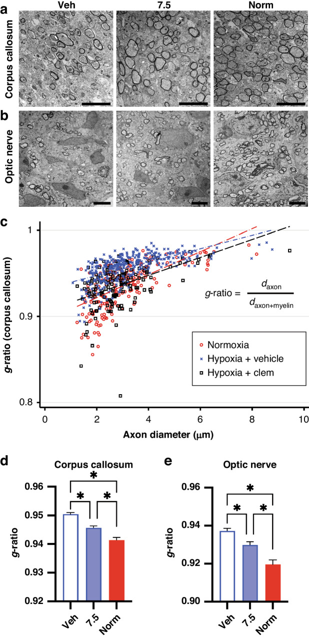
Fig. 4. Clemastine improves myelin thickness after chronic hypoxia exposure in neonatal mice.

a Representative electron microscopy images of the corpus callosum at 4700X magnification. Scale bars, 5 um. b Representative electron microscopy images of optic nerves at 2800X magnification. Scale bars, 5 um. c Scatter plot of g-ratios and axon diameters from the corpus callosum at P14, with superimposed linear regressions from normoxia-exposed (red), hypoxia-exposed/vehicle-treated (blue), and hypoxia-exposed/7.5 mg/kg/day clemastine-treated (black, “Hypoxia + Clem”) mice. daxon, diameter of axon; daxon+myelin, diameter of axon plus myelin sheath. d Bar plot comparing mean g-ratios in the corpus callosum. Error bars represent S.E.M. *p < 0.05, Kruskal–Wallis followed by post hoc Dunn’s tests. e Bar plot comparing mean g-ratios in optic nerves. Error bars represent S.E.M. *p < 0.05, Kruskal–Wallis followed by post hoc Dunn’s tests.
