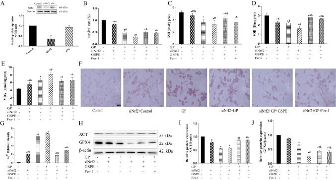
Fig. 6.
SiNrf2 exacerbates ferroptosis induced by glucolipotoxicity in MIN6 cells. A Nrf2 protein expression detected and quantified using western blot. B Cell viability measured in MIN6 cells after silencing Nrf2 using the CCK8 assay. C–E GSH, SOD, and MDA levels determined in MIN6 cells after silencing Nrf2 genes. F, G Lillie staining was used to detect and quantify ferrous iron content (blue) in MIN6 cells after silencing Nrf2 genes (× 400). H–J XCT and GPX4 protein expression levels detected and quantified using western blot analysis. Data are expressed as mean ± SD (*P < 0.05 vs. Control; #P < 0.05 vs. GP; &P < 0.05 vs. siNrf2 + GP)

