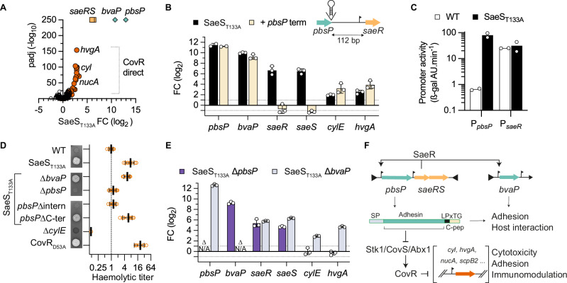
Fig. 4. Adhesin-dependent wiring of the SaeR and CovR regulatory networks.
A Volcano plot of significant fold change in the SaeST133A mutant. RNA-seq analyses with biological triplicate (n = 3) were analysed with Benjamini and Hochberg multiple comparisons to adjust P-values. Dot colours highlight the stratification of activated genes between pbsP and bvaP (green), the saeRS operon (orange) and the CovR-regulated (red) genes. B Indirect positive feedback loop of the saeRS operon. The pbsP and saeRS genes are separated by a 112 bp intergenic region containing a saeR transcriptional start site located 31 bp from the SaeR start codon. Transcriptional activation of pbsP and saeRS is uncoupled by the integration of a canonical terminator at the 3’ end of pbsP. Fold changes of selected genes are quantified by RT-qPCR in the SaeST133A (black bars) and in the SaeST133A + pbsP terminator (light bars) mutants. Bars represent the mean with SD of biological replicate (n = 3). C Activities of the PpbsP and PsaeR promoters in the WT and SaeST133A mutant. Bars represent the activity of the ectopic ß-galactosidase reporter system under the control of the tested promoters in the WT (white bars) and the SaeST133A (black bars) mutant. Bars represent the mean of two biological duplicates (n = 2). D Hyper-haemolytic activity of the SaeST133A mutant is dependent on the PbsP adhesin. Qualitative and semi-quantitative haemolytic activity is tested on Columbia blood agar media and with defibrinated horse blood, respectively. The ∆cylE and CovRD53A mutants are included as negative and positive controls, respectively. Haemolytic titres are normalised against the WT strain. Individual data points from biological replicates (n = 10, except for WT n = 9, CovRD53A n = 8, and ∆cylE n = 4) are represented with their mean +/– SD. E Upregulation of the PbsP adhesin activates CovR-regulated genes. Transcriptional fold change of selected genes by RT-qPCR in the SaeST133A ∆pbsP (light blue) and SaeST133A ∆bvaP (dark blue) double mutants. Bars represent the mean with SD of biological replicate (n = 3). F Wiring diagram of the SaeRS signalling pathway. The saeRS operon is transcribed at a basal level by a constitutive promoter. Upon TCS activation, the SaeR regulator activates the transcription of genes encoding the PbsP and BvaP virulence factors and indirectly its own operon through a pbsP terminator readthrough. The over-expression of the PbsP adhesin domain, but not the carboxy-terminal part containing the LPxTG anchoring motif and the hydrophobic C-peptide, is necessary to trigger CovR-regulated virulence factor expression via the Stk1/CovS/Abx1 regulatory proteins. Source data for panel (A) are provided in Supplementary Data 4 and as a Source Data file for panels (B, C, D, and E).

