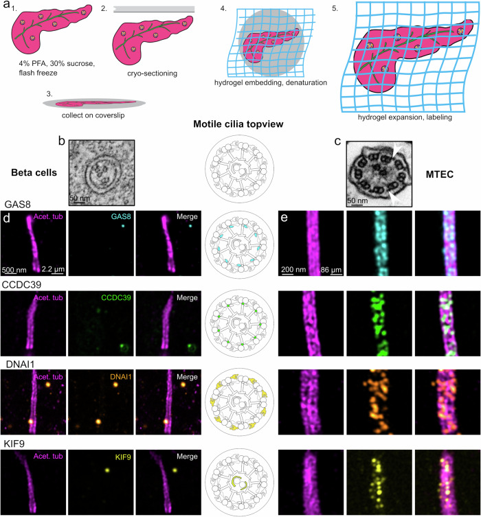
Fig. 3. Motility components do not localize to primary cilia of mouse beta cells.
a Schematic overview of pancreas U-ExM, including fixation, sectioning, collection, and expansion procedure. b Cross-sectional view of a beta cell primary cilium showing incomplete 9 + 0 organization. Scale bar: 50 nm. c Cross-sectional view of isolated mouse tracheal epithelium, displaying classic 9 + 2 organization. White arrows denote example outer dynein arms. Scale bar: 50 nm. d U-ExM images of beta cell cilia. Scale bar left, expansion factor corrected: 500 nm. Scale bar right, true scale: 2.2 µm. e U-ExM images of isolated mouse tracheal epithelium cilia (MTEC). Scale bar left, expansion factor corrected: 200 nm. Scale bar right, true scale, 0.86 µm. Cartoon indicates motility component localization in motile cilia. Component localization is determined by acetylated tubulin (magenta), GAS8 (cyan), CCDC39 (green), DNAI1 (hot orange), and KIF9 (yellow). U-ExM data were obtained from 2 individual mice.

