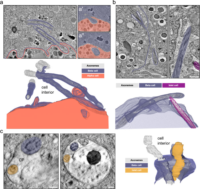
Fig. 7. Cilia-cell and cilia-cilia interaction.
a Single tomographic slices of primary cilia in human islets from one donor. There are 3 cilia originating in a beta cell with the basal body of one cilium (1) and the axonemes of 2 other cilia visible in the slice (2 and 3, purple). The neighboring alpha cell is outlined in orange. In magnified views of distant slices through the tomogram (a’ and a”) the end of cilium 2 is visible protruding and extending into a neighboring alpha cell (orange). Axonemes, basal bodies, and centrioles are rendered in gray. Scale bars: 500 nm and 100 nm. b Single tomographic slices of mouse beta cells of one isolated mouse pancreatic islet with 2 primary cilia (purple and magenta) originating from a beta cell (purple) and another islet cell (magenta) meeting in the space between cells. The side view shows the axonemes of the cilia in close proximity. The 3D view shows the segmentation of ciliary and plasma membranes together with the axonemes. Scale bars: 200 nm. c Tomographic slices of a ssET volume of one mouse pancreatic islet showing a large ciliary pocket shared by a primary cilium originating from a beta cell (purple) and a primary cilium of unknown origin (yellow). The 3D rendering shows the interaction of the two cilia. Axonemes are in gray. Scale bars: 100 nm.

