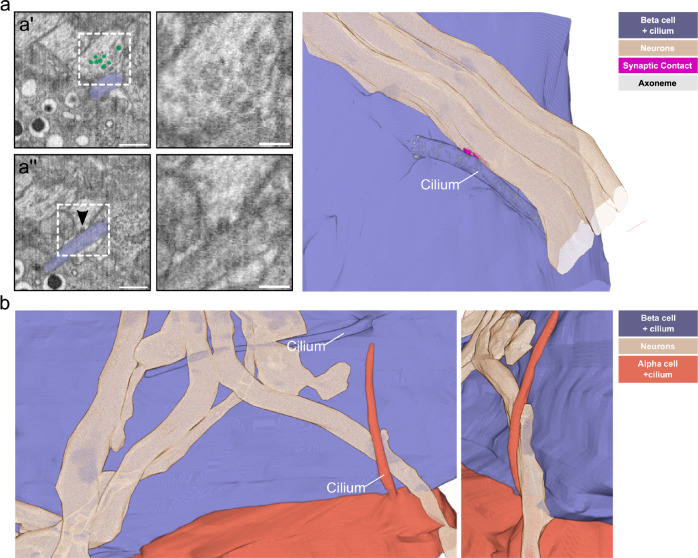
Fig. 8. Islet cell primary cilia-neuron connections.
a A single slice of a FIB-SEM volume of one P7 mouse pancreas sample with an axon and a beta cell with a primary cilium (purple) in contact with each other. Scale bar: 1 µm. In a’, synaptic vesicles close to the cilium-neuron contact are highlighted in green. Scale bar: 500 nm. The boxed area is shown magnified in the right panel without highlighting the vesicles. Scale bar: 100 nm. The arrowhead in a” points to a synaptic vesicle likely undergoing exocytosis towards the cilium. Scale bar: 500 nm. The magnified image in the right panel shows the area of the fusion event. Scale bar: 100 nm. The 3D rendering shows the contact of a primary cilium (purple) with one of the axons (beige), including a segmentation of the synaptic density (magenta). b 3D rendering of primary cilia from a beta (purple) and an alpha cell (orange) in contact with the same axon (beige). The smaller image shows a tilted magnified view of the alpha cell cilium-axon interaction.

