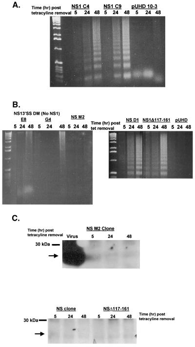
FIG. 3.
The RNA-binding domain of NS1 is required for apoptosis. (A) Confluent cultures of MDCK cells expressing pUHD 10-3 containing the NS1 gene from A/Udom/72 or empty vector (pUHD) were washed two times with PBS and then incubated for 5, 24, or 48 h in anhydrotetracycline-free MEM containing 1% FBS at 37°C in 5% CO2. DNA was collected and analyzed for DNA fragmentation by agarose gel analysis. Two different clones, C4 and C9, expressing NS1 protein were analyzed. (B) Confluent cultures of MDCK cells expressing pUHD 10-3 containing an NS mutant that fails to produce NS1 protein (NS13′SS DM clones E8 and G4), a mutation in the RNA-binding domain (M2), full-length NS (clone D1), or a deletion of the effector domain (NS1Δ117-161) were washed two times with PBS and then incubated for 5, 24, or 48 h in anhydrotetracycline-free MEM containing 1% FBS at 37°C in 5% CO2. DNA was collected and analyzed for DNA fragmentation by agarose gel analysis. (C) Confluent cultures of MDCK cells expressing pUHD 10-3 containing the NS M2 (RNA-binding domain mutation), full-length NS gene from A/Ty/Ont/66, or NS1Δ117-161 were washed two times with PBS and then incubated for 5, 24, or 48 h in medium without anhydrotetracycline. Cell monolayers were lysed, and 5 mg of total protein was loaded per lane under reducing conditions and resolved by SDS-PAGE. After being transferred to nitrocellulose, proteins were probed for NS with a rabbit polyclonal antibody against NS. Bands were detected by enhanced chemiluminescence as instructed by the manufacturer. The arrow indicates the location of NS. Molecular size markers are indicated on the left. MDCK cells infected with A/Ty/Ont/66 at an MOI of 0.5 for 24 h served as a positive control (virus lane).

