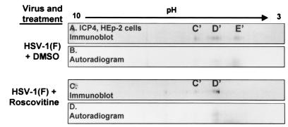
FIG. 5.
Immunoblots (A and C) and corresponding autoradiograms (B and D) of ICP4 in two-dimensionally separated 32P-orthophosphate-labeled HSV-1-infected HEp-2 cell lysates to determine roscovitine-sensitive ICP4 phosphorylation. HSV-1-infected HEp-2 cells were treated with roscovitine or an equivalent volume of DMSO at 5 h after infection in phosphate-free medium. Infected cells were labeled with 32P-orthophosphate from 6 to 10 h after infection in the presence of roscovitine. Cells were harvested at 10 h postinfection, and whole-cell lysates were subjected to two-dimensional electrophoresis. Membranes were developed by autoradiography and immunoblotted with an ICP4 antibody. Isoform designations are idential to those shown in Fig. 3.

