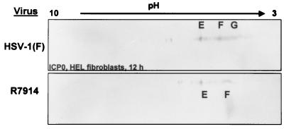
FIG. 8.
Immunoblots of ICP0 in two-dimensionally separated lysates of HSV-1 or R7914 (alanine substituted for aspartic acid of ICP0)-infected HEL fibroblasts. The cells were harvested at 12 h after infection, lysed, and subjected to two-dimensional electrophoresis. Immunoblots were reacted with anti ICP0 antibody. Isoform designations are the same as in Fig. 2.

