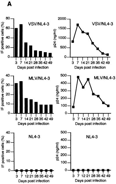
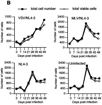
FIG. 2.
Expression of HIV-1 antigens, viability, and growth kinetics in fetal astrocytes infected with native and pseudotyped HIV-1. (A) Astrocyte cultures were infected with VSV/NL4-3, MLV/NL4-3, or NL4-3 as indicated and monitored for HIV-1-specific antigens expression by IF staining with AIDS sera and fluorescein isothiocyarate-conjugated second antibody (left panel) or by production of p24 in cell supernatants (right panel). The proportion of IF-positive cells was determined by counting at least 200 cells each in three different fields under ×20 magnification, using an Olympus BH-2 fluorescence microscope. (B) At the indicated times after infection, total cell numbers and total viable-cell numbers per system (×1,000) were determined as described in Materials and Methods.


