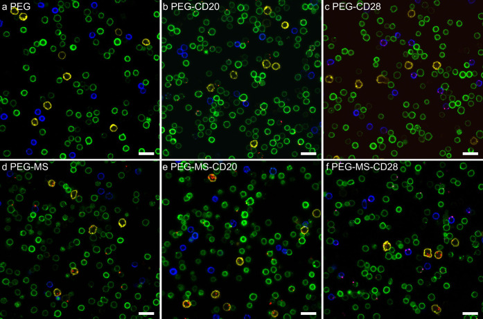
Figure 4.
Confocal microscopy images showing the association of BsAb-functionalized PEG or PEG-MS nanoparticles with blood cells from a CLL patient. (a–c) PEG and (d–f) PEG-MS nanoparticles functionalized with or without anti-PEG/anti-CD20 or anti-PEG/anti-CD28 BsAbs were incubated with PBMCs from a CLL patient for 1 h at 37 °C, followed by phenotyping cells with antibody cocktails and imaging by confocal microscopy. Green, CD20+ CLL cells; blue, CD3+ T cells; yellow, CD14+ monocytes; red, fluorescence-labeled PEG or PEG-MS nanoparticles. Scale bars are 20 μm.

