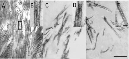
Figure 3.
Ultrastructural examination of microtubule bundles induced by AtMAP65 proteins. A and B, Large microtubule bundles induced by AtMAP65-1. Note that microtubules were evenly spaced out. The insert (B) showed ladder-like cross-bridges between adjacent microtubules. C, D, E, and F, Microtubule mesh-like network induced by AtMAP65-6. A few microtubules were found to be linked together, and microtubules were generally scattered (C and E). Inserts (D and F) show cross-bridges formed between microtubules. Scale bar = 0.5 μm in A, C, and E, and 0.05 μm in B, D, and F.

