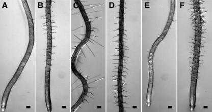
Figure 2.
Epidermal CFR phenotypes of wvd6 and rhd3 roots. Seedlings were grown on a tilted agar surface and photographed 9 DAG. Root images were taken either at the tip (A, B, E, and F) or at a position within the mature zone where root hair length was maximal (C and D). A and C, Wild-type Ws; B and D, wvd6; E, wild-type No-0; and F, rhd3-3. Scale bars = 100 μm.

