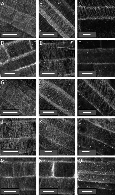
Figure 7.
Organization of the cortical microtubule network in wild-type and mutant root tips. Seedlings grown on vertical agar surfaces were subjected to the immunolocalization protocol described in “Materials and Methods” and stained with mouse antitubulin antibodies. Micrographs were taken in the middle of the distal elongation zone (meristem; left column, A, D, G, J, and M), in the middle of the central elongation zone (central column, B, E, H, K, and N), or at the maturing zone where root hairs initiate (right column, C, F, I, L, and O) of wild-type Col (A–C), rhd3-1 (D–F), cob-1 (G–I), erh2-1 (J–L), and eto1-1 (M–O) 5-d-old seedlings. White bars = 10 μm.

