Figure 1.
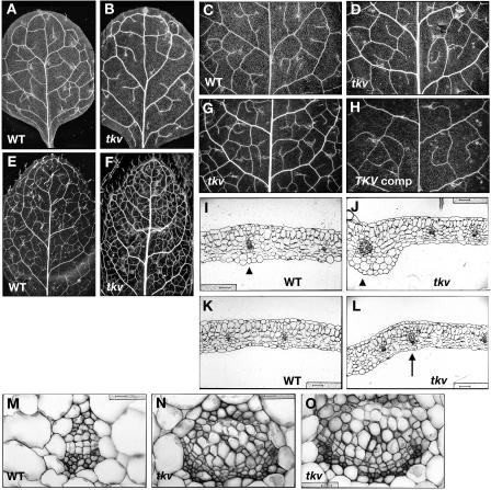
Increased vein thickness and vascularization in tkv leaves. A to H, Cleared leaves of wild type (A, C, and E), tkv (B, D, F, and G), and fully restored tkv containing wild-type copy of the ACL5 transgene (H) were viewed under dark-field illumination to visualize the venation pattern. A and B, First pair of juvenile leaves. C, D, G and H, Second pair of juvenile leaves. E and F, Adult leaves. All leaves are in same magnification, and leaves used for comparison are developmentally equivalent (have similar number of secondary veins that form prominent arches that begin and end at the midvein) and representative of numerous samples. I to O, Transverse sections through the midvein (I, J, and M–O) and lateral veins (K and L) of fully expanded juvenile leaves. I, K, and M, Wild type. J, L, N, and O, tkv. Arrowheads indicate the midvein, and arrow indicates a secondary vein. In wild type, aside from the midvein, higher-order veins are indistinguishable by vein thickness. Scale bars in I to L = 50 μm, and scale bars in M to O = 16 μm.

