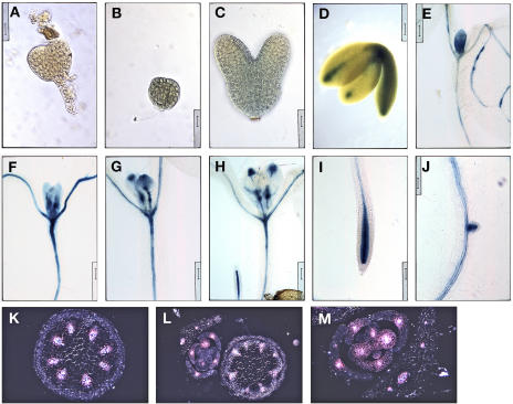
Figure 9.
ACL5 promoter activity is associated with provascular/procambial cells. A, Nontransgenic globular-staged embryo was stained for GUS activity. Note the absence of GUS stain. Scale bar = 16 μm. B to M, Wild-type plants containing a transgenic copy of the ACL5 promoter-driven GUS reporter gene were stained for GUS activity and viewed under bright-field illumination unless noted otherwise. B, Globular-staged embryo. Scale bar = 16 μm. C, Torpedo-staged embryo. Staining is weak and diffuse. Scale bar = 16 μm. D, Bent cotyledon-staged embryo. Scale bar = 62.5 μm. E to J, Cleared GUS-stained week-old seedlings were arranged in developmental order. Note that GUS stain gradually localizes to and around PC cells. Scale bars = 100 μm. K to M, Transverse sections through inflorescence stems and axillary buds were viewed under dark-field illumination to visualize GUS staining, which appears pink. Note that GUS staining is localized to developing vasculature. Scale bars = 50, 100, and 50 μm, respectively.

