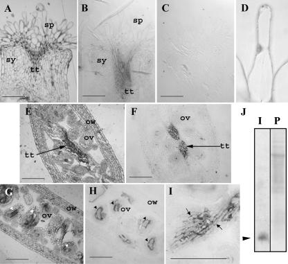
Figure 2.
Plantacyanin is localized to the wild-type Arabidopsis stigma and to the transmitting tract tissues of the style and ovary. Toluidine blue-stained sections after immunolocalization (A, E, G) show the tissue structures of the Arabidopsis pistil. Immunolocalizations (B–D, F, H, and I), using an antibody against recombinant plantacyanin made in E. coli, show that the transmitting tract and the embryo sacs produce plantacyanin protein. No signal was found when the preimmune serum was incubated with the section (C). The expression of Arabidopsis plantacyanin in the stigmatic papilla cell was detected at a very low level on the LR White-embedded sections (fixation with FAA; B), and at a slightly higher level using glutaraldehyde fixation (D). Expression in the stylar transmitting tract is high (B). Ovary transmitting tract tissues (the septum; F) and the embryo sacs show strong localization as well (arrowheads in G and H, longitudinal sections at flower stage 12). An enlarged image of the septum transmitting tract cells (I) shows the extracellular localization of plantacyanin (arrows). The antibody used for immunolocalization recognized a single plantacyanin band (arrowhead in J) on an immunoblot analysis (labeled as I in J) of total cell proteins extracted from Arabidopsis inflorescences; the blot was stained with Ponceau S (labeled as P in J) before immunoblotting. sp, Stigmatic papilla cell; sy, style; tt, transmitting track; ov, ovule; ow, ovary wall. Bars = 100 μm.

