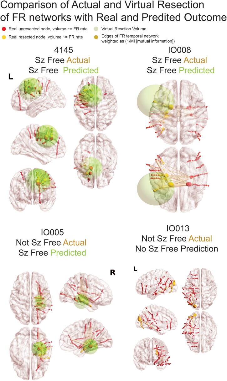
Figure 5.
Illustration of FR networks and real and virtual resections. In the four patients, the sizes of red (unresected) and yellow (resected) nodes (i.e. SEEG electrode contacts) are proportional to the relative FR rate. The edges (pale yellow), connecting the nodes to one another, are weighted in size by the inverse of the MI of FR temporal correlations between the two nodes. The green sphere denotes the borders of the virtual resection. The centre of the sphere is the node with most FR autonomy and/or highest FR rate and has a margin of 1 cm. The four FR metrics (FR RR, spatial FR net and temporal FR net-A, B) are derived from comparison of the sets of FR-generating contacts in and outside of the virtual resection sphere, and these factors are used by the SVM to predict virtual seizure (sz) freedom. If the SVM predicts non-seizure (non-sz) freedom, the virtual resection model iterates, and the virtual resection sphere may expand depending on whether the spatial location of the next top node that generates FR at higher rates and most autonomy is outside the sphere. In the case that the sphere expands, the new margins are extended by 1 cm. If the virtual resection includes three lobes, it is considered a failure. As shown, extension of the virtual resection sphere outside of the spatially sampled regions, and outside the brain, does not increase the number of nodes in the virtual resection set. Contralateral nodes within the virtual resection sphere are also excluded from the virtual resection set of nodes. For patients 4145 and IO008, who were rendered seizure free, the virtual resection that was predicted as sufficient for virtual seizure freedom included a set of nodes that partially overlaps with the set of resected nodes. In patient 4145, the contacts in the virtually resected set (red and yellow nodes within green sphere) were larger than the set of resected nodes (yellow nodes). In patient IO008, the difference between the set of nodes in the virtual resection and actual resection was smaller (see Table 3). Patient IO005 was not seizure free, but the virtual resection that predicted a seizure-free outcome was more posterior and included nodes with high FR rate and low MI edges. Patient IO013 was not seizure free, but the virtual resection did not produce seizure freedom because it required a resection of the occipital, parietal and temporal lobe.

