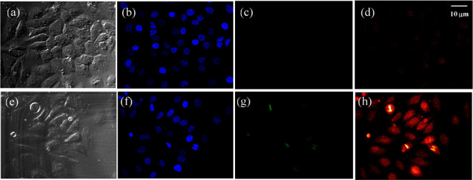
Figure 3.

Confocal fluorescence images of HeLa cells excited with a 488 nm laser. The images were collected at bright field (a,e), DAPI (b,f), green channel (c,g), and NIR channel (d,h: 700–800 nm). Top (control): Cells were incubated with Zinhbo-9 for 60 min at 37 °C in Dulbecco’s modified Eagle’s medium (DMEM). Bottom: Cells were first treated with Zn2+ (10 μM) for 30 min and further exposed to Zinhbo-9 (10 μM) for another 60 min at 37 °C in DMEM.
