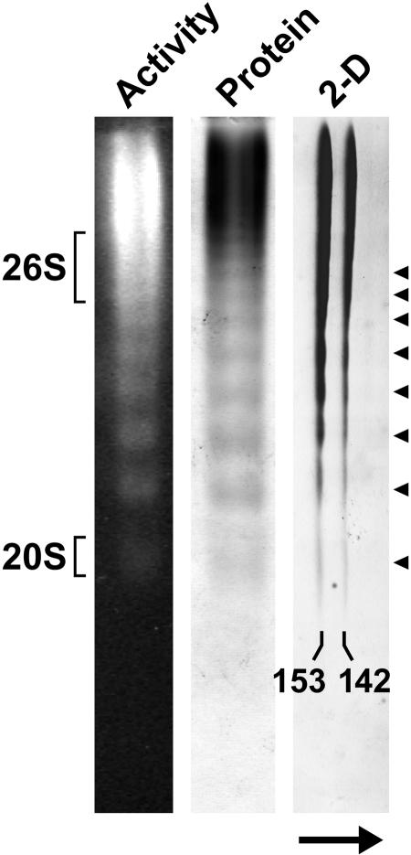
Figure 2.
Native PAGE of Arabidopsis TPPII. The peak fraction from the Superose HR6 FPLC was subjected to native PAGE and TPPII was either detected by fluorescence following overlay of the gel with the substrate AAF-AMC or by staining with silver nitrate. Migration positions of the 26S proteasome and its 20S CP in an adjacent lane are indicated. For two-dimensional PAGE, TPPII was first subjected to native PAGE followed by SDS-PAGE in the second dimension and the gel was then stained with silver nitrate. The arrow indicates the electrophoretic direction of the SDS-PAGE. The positions of the 153- and 142-kD forms of TPPII are shown. Arrowheads locate the series of discrete size species of TPPII observed by native PAGE.

