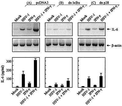
FIG. 7.
Induction of IL-6 by HSV-1 in macrophages stably transfected with dominant negative IκBα and p38. RAW 264.7-derived cell lines with the following characteristics were generated: pcDNA3 (A), dominant negative IκBα (B), dominant negative p38V (C). The cells were seeded, infected with 3 × 10⁶ PFU of HSV-1 (KOS) per ml, and stimulated with 10 IU of IFN-γ per ml. After 4 h, cells to be analyzed for mRNA expression were lysed and total RNA was extracted. IL-6 and β-actin were detected by RT-PCR using primers specific for the two mRNA species (upper panel). Other cells were left for 24 h, at which point supernatants were harvested and analyzed for IL-6 protein by ELISA. The results (lower panel) are shown as means ± standard errors of the means.

