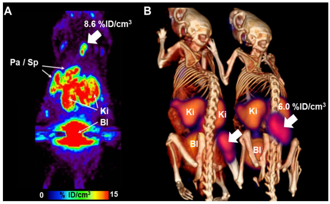
Figure 15.
[18F]FASu PET imaging in mice bearing SKOV-3 and EL4 xenograft tumors. (A) The image shows the maximum-intensity projection of SKOV-3 tumor-bearing nude mice. (B) PET/CT image summed over 110–120 min after injection in Rag2 M mice bearing EL4 xenograft tumor. The tumor is indicated by a white arrow. PET/CT, positron emission tomography/computed tomography; Pa, pancreas; Sp, Spleen; Ki, Kidney; Bl, Bladder. Reproduced from [52], copyright © 2014, Journal of Nuclear Medicine.

